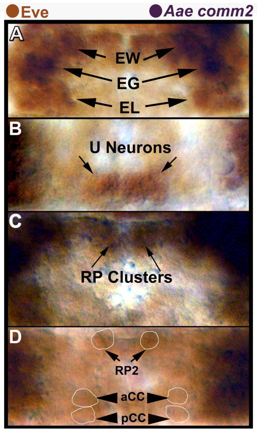
Fig. 4. Lack of Aae comm2 expression in neurons that do not cross the midline.
The EL (A) and U (B) neuron clusters, RP2, aCC, and pCC neurons (D) are marked by expression of Eve protein (brown). These Eve-positive neurons do not express Aae comm2 mRNA (dark purple, A–D), and their axons do not cross the midline. The EG, EW (A) and RP neuron (RP1, 3, 4; RP2 is out of focus) clusters (C) were positionally identified with respect to Eve-positive neurons; these neurons (with the exception of RP2) project axons to the midline and express Aae comm2 (dark purple). All images are oriented anterior upward and correspond to different segments/focal planes of the nerve cord from a single filleted 51 hour old embryo. Ventral focal planes of the nerve cord are shown in A and B, while C shows a more dorsal plane of focus. Panel D shows the dorsal-most plane of focus in this nerve cord. Light Eve staining permitted examination of comm2 expression in Eve-positive neurons of A. aegypti embryos in which tissue autofluorescence hinders fluorescent immunohistochemistry. Lightly-stained Eve-positive cell bodies were circled in some panels (D) to facilitate interpretation of these data.

