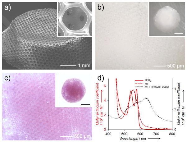
Figure 1.
a) A representative SEM image showing a PLGA inverse opal scaffold with uniform pores and a long-range ordered structure. The inset shows a magnified view of a pore on the surface, revealing the uniform windows connecting to the pores underneath. Scale bar: 100 μm. b, c) Optical micrographs showing (b) a pristine PLGA inverse opal scaffold and (c) a PLGA inverse opal scaffold after doping with MTT formazan to render it purple in color. d) UV-vis extinction spectra of hemoglobin, deoxy-hemoglobin, and MTT formazan crystals. Formazan has an absorption peak at approximately 650 nm, while blood does not show strong absorption at >600 nm.

