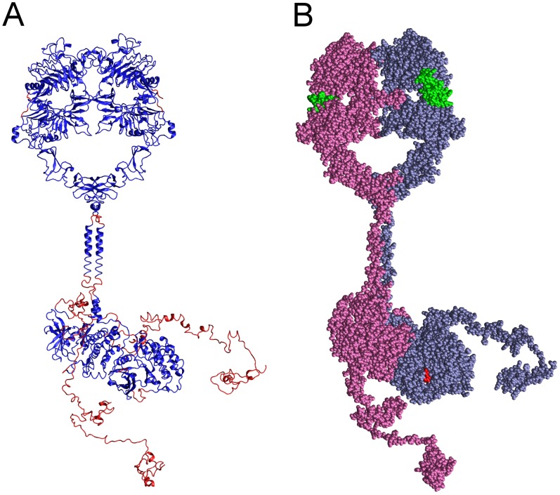
Figure 1. Complete structural model of the dimeric EGFR.
Shown in two representations is the complete structural model of a dimer of EGFR molecules, each with a bound EGF molecule and bound AMPPNP.2Mg2+ substrate complex, generated herein. (A) Backbone conformation and secondary structure of the modeled EGFR polypeptides, with segments derived from published crystallographic structures colored blue and modeled segments colored red. (B) Van der Waals representation of the EGFR dimer, with the monomers having active (receiver) and inactive (activator) conformation kinases colored ice-blue and mauve, respectively, bound EGF molecules colored green, and bound AMPPNP.2Mg2+ substrate complexes colored red.

