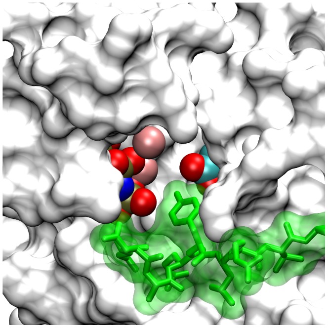
Figure 2. Active conformation PTK domain with bound nucleotide and peptide substrates in the complete EGFR model.

Shown in an accessible surface representation are residues of the active conformation PTK domain (white) with its catalytic Asp-813 and bound AMPPNP.2Mg2+ substrate complex highlighted in CPK coloring. The docked nine-amino acid P-site-1173 peptide (sequence ENAEYLRVA) is shown in green. Note that the tyrosine hydroxyl of the peptide substrate is in close proximity to both the carboxyl group of Asp-813 and the γ-phosphate of the AMPPNP substrate analog. Similar models were generated with each of the eight distinct P-site peptides (see Table 1) docked in the active site.
