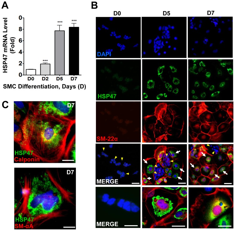
Figure 1. HSP47 is expressed during SMC differentiation of ESC.
A–C, HSP47 is increased during ESC-SMC differentiation. Murine ESC were differentiated into SMC in the presence of Collagen IV for the time indicated, followed by quantitative RT-PCR analysis of HSP47 mRNA (A), immunofluorescent staining with HSP47 (green) and SM-22α (red) antibodies (B), calponin (red) and SM-αA (red) (C). DAPI was included to counterstain the nucleus (blue). Scale bar, 30 µm. Images shown are representative of at least three separate experiments, whereas graphs are shown as mean ± SEM of at least three independent experiments, ***P<0.005 compared with Day 0.

