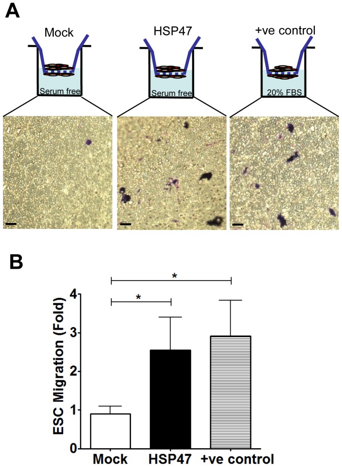
Figure 3. HSP47 over-expression induces ESC chemotaxis.
A and B, over-expression of HSP47 induces chemotaxis of ESC. ESCs were transfected with pCMV6-HSP47 plasmid (1 µg/1×106 cells) via electroporation and subjected to 5.0micron transwell chemotaxis assays 48 hours later. Chemotaxis of ESCs (either HSP47 or mock) towards serum free media in the transwells was documented following 1% crystal violet staining. (Scale bars, 30 µm) Chemotaxis of ESCs towards media containing 20% FBS was used as a positive control. Chemotaxis index was defined as the mean number of ESCs counted per 10 random fields of view with 20× objective and presented as fold increase compared to the mock control. Graphs are shown as mean ± SEM of three independent experiments. *p<0.05 compared with mock control.

