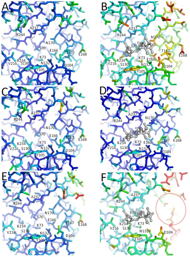
Figure 8. Protein disorder induced by SA2-13 binding.
Temperature factors of protein atoms are represented by rainbow color-ramping from blue to red for temperature factors ranging from <5 to >40 Å2. Depicted are the structures of K234R SHV (A) and (B), wt SHV-1 (C) and (D), and R164S SHV (E) and (F). Uncomplexed structure are shown on the left (A), (C), and (E), whereas SA2-13 bound structures are on the right (B), (D), (F) with SA2-13 being depicted in grey ball-and-stick model. Completely disordered region not modeled in the SA2-13 bound R164S SHV structure is highlighted by red dashed oval.

