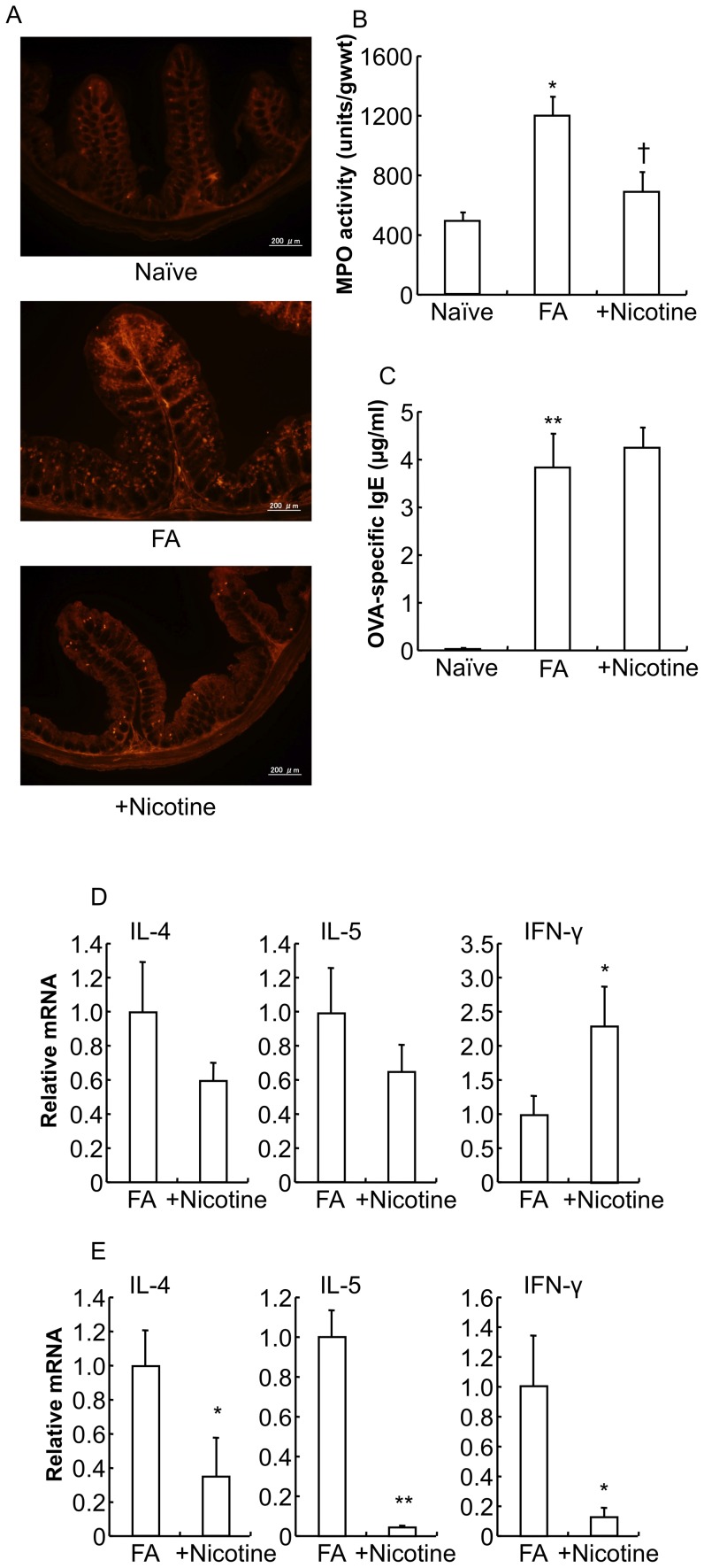
Figure 9. Effect of nicotine on the pathology of food allergy in mice.
A: The proximal colons of naïve mice, FA mice, and mice treated with the subcutaneous administration of nicotine were stained with mMCP-1 antibodies. Scale bars represent 200 µm. B: MPO activity was measured in the colon of naïve mice, FA mice and nicotine-treated FA mice. *P<0.01 vs. naïve mice. †P<0.05 vs. FA mice (n = 3–10 mice per group). C: The level of OVA-specific IgE in the plasma of naïve mice, FA mice and nicotine-treated FA mice is shown. The level of OVA-specific IgE in the plasma was determined using an ELISA kit. **P<0.01 vs. naïve mice (n = 5 mice per group). IL-4, IL-5 and IFN-γ cytokine mRNA expression in the spleen (D) and proximal colon (E) from FA mice and nicotine-treated FA mice were measured by real-time PCR. Relative mRNA levels of cytokines were normalized to GAPDH expression. *P<0.05, **P<0.01 vs. FA mice (n = 3–6 mice per group).

