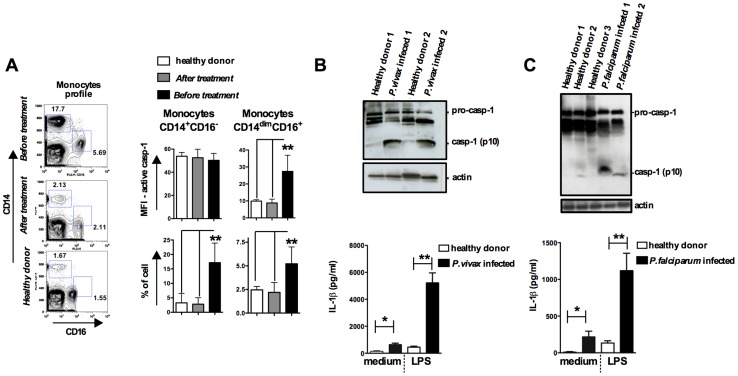
Figure 5. Monocytes are the major source of active caspase-1 during malaria.
PBMCs were obtained from either P. vivax or P. falciparum malaria patients as well as from healthy donors. (A) The gate was set based on monocytes profile in PBMCs from P. vivax infected patients. PBMCs were stained with anti-CD14 and anti-CD16 antibodies to determine the presence of different monocyte subsets. These cells were gated based on FSC and SSC to avoid neutrophil contamination. When the CD14dimCD16+ gate was moved down in PBMCs from healthy donors or from malaria patients after treatment, we did not detect any active caspase-1. The bar graphs show the flow cytometry analysis of PBMCs from five P. vivax infected subjects before and after malaria treatment primaquine and chloroquine, as well as eight healthy donors. To determine the median fluorescence intensity (MFI) and frequency of (CD14+CD16−) and (CD14dimCD16+) that are active caspase-1, we used the FLICA assay. (B – top panel) Active caspase-1 (p10) was detected in lysates of PBMCs from P. vivax or (C – top panel) P. falciparum infected individuals by Western blot. (B and C – bottom panel) PBMCs were stimulated with LPS (100 ng/ml) for 24 hours, and levels of IL-1β assessed in the culture supernatants by ELISA. Significant differences are *p<0.05 and **p<0.005 as indicated by the unpaired t test with Welch correction or Mann-Whitney test when a normality test failed.

