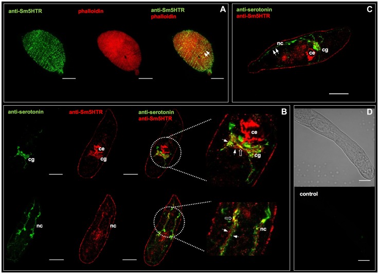
Figure 3. Immunolocalization of Sm5HTR in schistosomulae.
In vitro transformed schistosomulae (3–8 days old) were probed with affinity-purified anti-Sm5HTR antibody and visualized by confocal microscopy. (A) Typical schistosomulum showing Sm5HTR (green) immunoreactivity in varicose nerve fibers along the length of the body (left panel). Animals were co-labelled with TRITC-phalloidin to visualize the musculature (middle panel) and the overlay (right panel) shows punctate bright yellow fluorescence, indicating sites of apparent co-localization between Sm5HTR and the outer circular muscles of the body wall (arrows). (B) Schistosomulae were co-labelled with anti-serotonin antibody (green) and anti-Sm5HTR antibody (red). The overlay shows close proximity between the two signals (solid arrows) in the cerebral ganglia (cg) and the main longitudinal nerve cords (nc). Sites of apparent co-localization are marked by open arrows. Sm5HTR immunoreactivity is also prominent in the developing caecum (ce). (C) Schistosomulum co-labelled with anti-serotonin antibody (green) and anti-Sm5HTR antibody (red). Lateral serotonin-containing nerve fibers (solid arrows) can be seen in close proximity to Sm5HTR immunoreactivity in the body wall region of the larva. cg, cerebral ganglia; ce, caecum; nc, nerve cord. (D) Transmission light and corresponding fluorescence image of a typical negative control. No significant fluorescence could be seen in any of the negative controls probed with secondary antibody only or antigen-preadsorbed antibody. Scale bars, 25 µm.

