
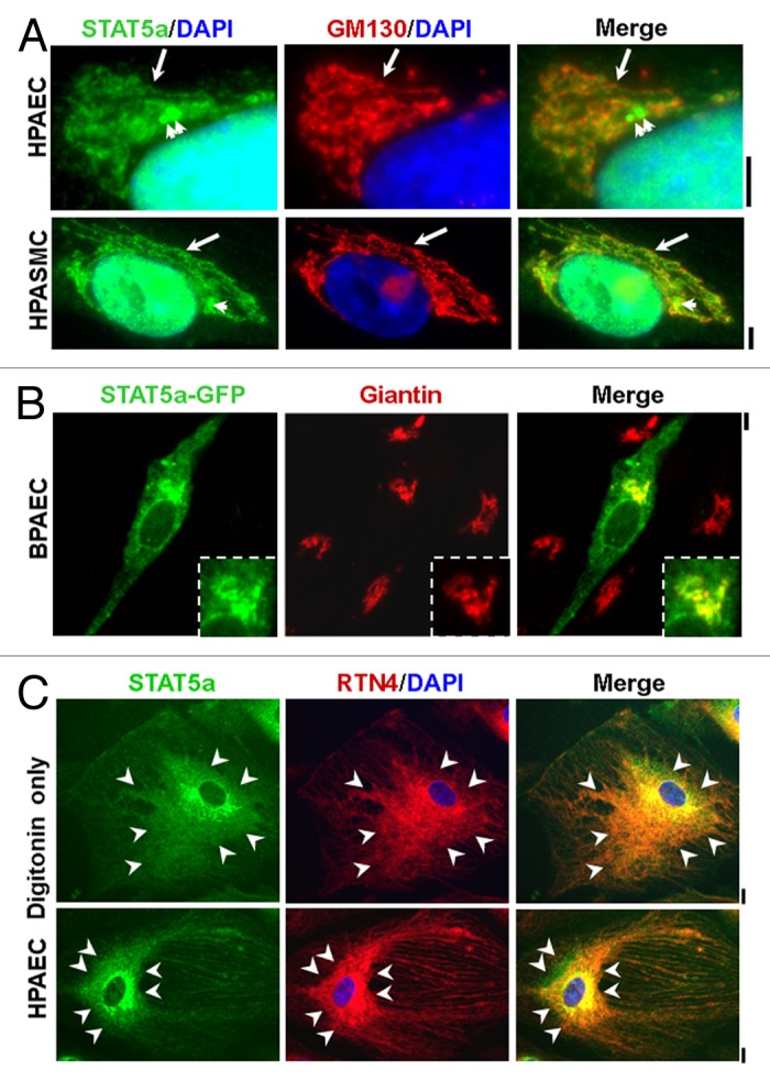
Figure 1. Association of STAT5A with the Golgi apparatus, endoplasmic reticulum and centrosomes. (A) Immunofluorescence imaging of the association of STAT5A with the Golgi tether GM130 (long arrow) and centrosomes (short arrows) in human pulmonary arterial endothelial (HPAEC) and smooth muscle cells (HPASMC) after fixation and then Triton permeabilization. Scale bars = 5 µm. (B) Fluoresence imaging of bovine PAECs transfected with a human STAT5A-GFP expression vector after fixation together with immunofluorescence imaging of the Golgi tether giantin. Scale bar = 5 µm; inset shows Golgi region at higher magnification. (C) Immunofluorescence colocalization of STAT5A with the endoplasmic reticulum (ER) structural protein RTN4/Nogo-B in HPAECs after fixation and permeabilization with only digitonin. Arrowheads demarcate the region of ER sheets; Scale bars = 10 µm. Illustrations in this composite figure were adapted from references 13 and 14.