
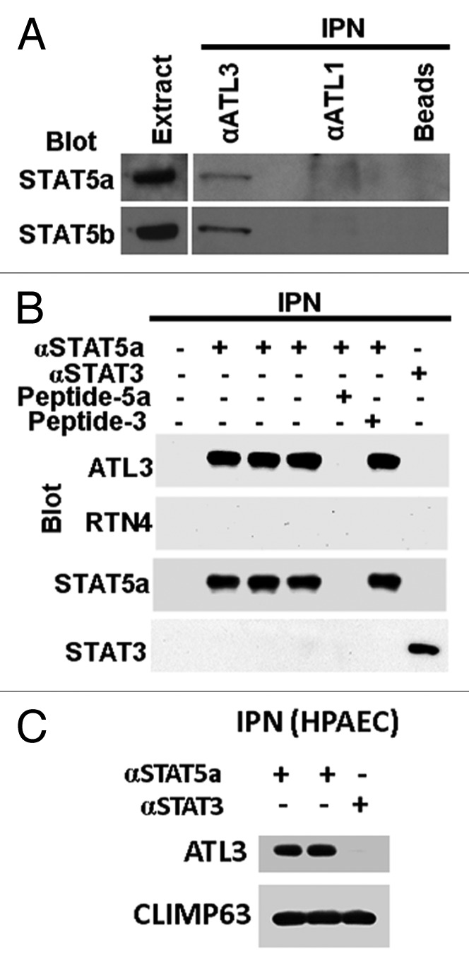
Figure 4. Association between STAT5A and STAT5B with ATL3 but not RTN4 in magnetic-bead cross-immunopanning assays and western blotting. (A) Immunopanning (IPN) of proteins from extracts of human endothelial cells (EA.hy926) was performed using anti-ATL3 or anti-ATL1 rabbit pAb as well as control beads alone (endothelial cells do not express ATL1; thus this also acts as a “negative” IgG control). The immunoisolates were western blotted for STAT5A or STAT5B as indicated. (B) Immunopanning was performed using extracts from human endothelial cells (EA.hy926 cells) using anti-STAT5A rabbit pAb in triplicate together with controls which included absence of any IgG, use of an irrelevant pAb (anti-STAT3 pAb) and prior incubation of the anti-STAT5A pAb with either relevant competing peptide (Peptide-5A) or an irrelevant competing peptide (Peptide-3). Western blotting of the immunoisolated complexes was performed as indicated. (C) Equal aliquots of HPAEC extracts were subjected to magnetic-bead immunopanning using anti-STAT5A or anti-STAT3 pAb. The immunoisolates were sequentially western blotted using Abs to ATL3 or CLIMP63 as indicated. Illustrations in this composite figure were adapted from references 13 and 14.