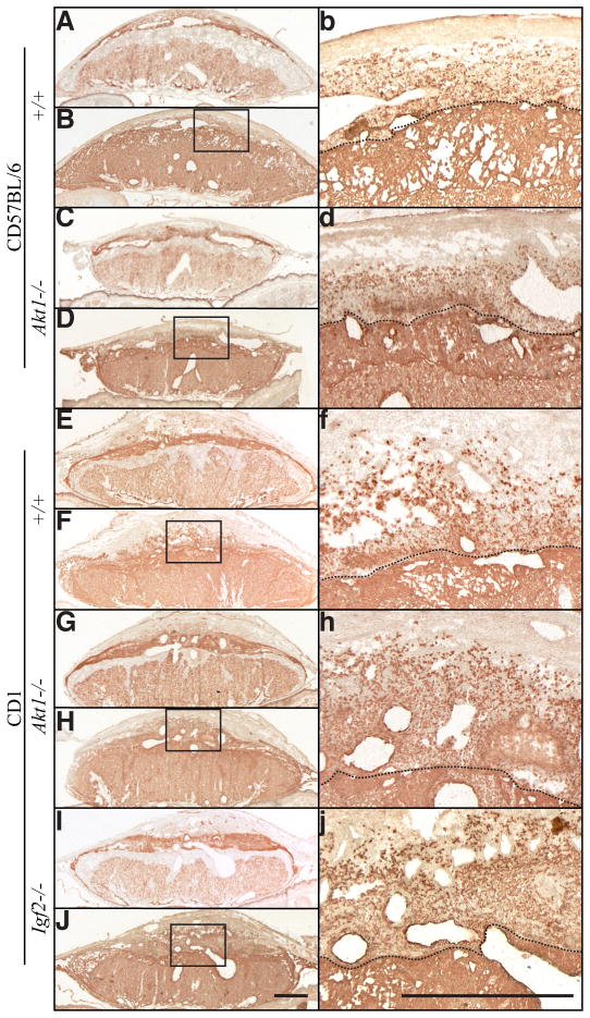
Fig. 4. Organization of the placentation site in wild-type and Akt1 and Igf2 nulls.
Isolectin B4 binding (A,C,E,G,I) and cytokeratin immunocytochemistry (B,D,F,H,J) were performed on placentation sites from gestation day 17.5 pregnant wild-type and Akt1 nulls on C57BL/6 and CD1 genetic backgrounds. Similar analyses were performed on CD1 Igf2 nulls. High magnification of the boxed areas in (B,D,F,H,J) are shown in images labeled with the respective lower case letters. The dashed line indicates the decidua-junctional zone interface. Scale bars, 1 mm.

