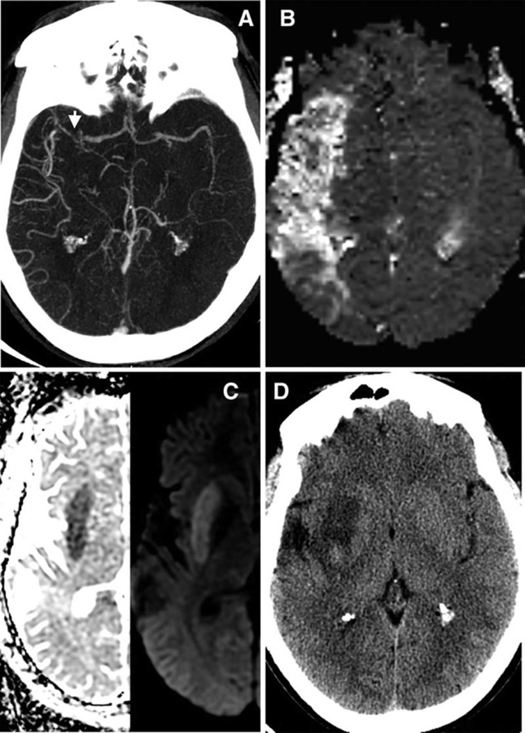
Figure 2. Example of a Low PIRI-score without significant mismatch loss.
34 year-old man presenting five hours after right hemispheric stroke onset with admission NIHSS score of 11. Axial images show: (A) acute proximal MCA occlusion with high collateral flow (versus contralateral) on CTA (arrow), (B) with a large MR-MTT hypoperfused lesion. (C) However normal insula on admission ADC/DWI (PIRI score 0,) and (D) only minimal (13%) tissue-at-risk infarcted on 48-hour follow-up CT despite lack of treatment with IV-tPA or IAT.

