FIGURE 2.
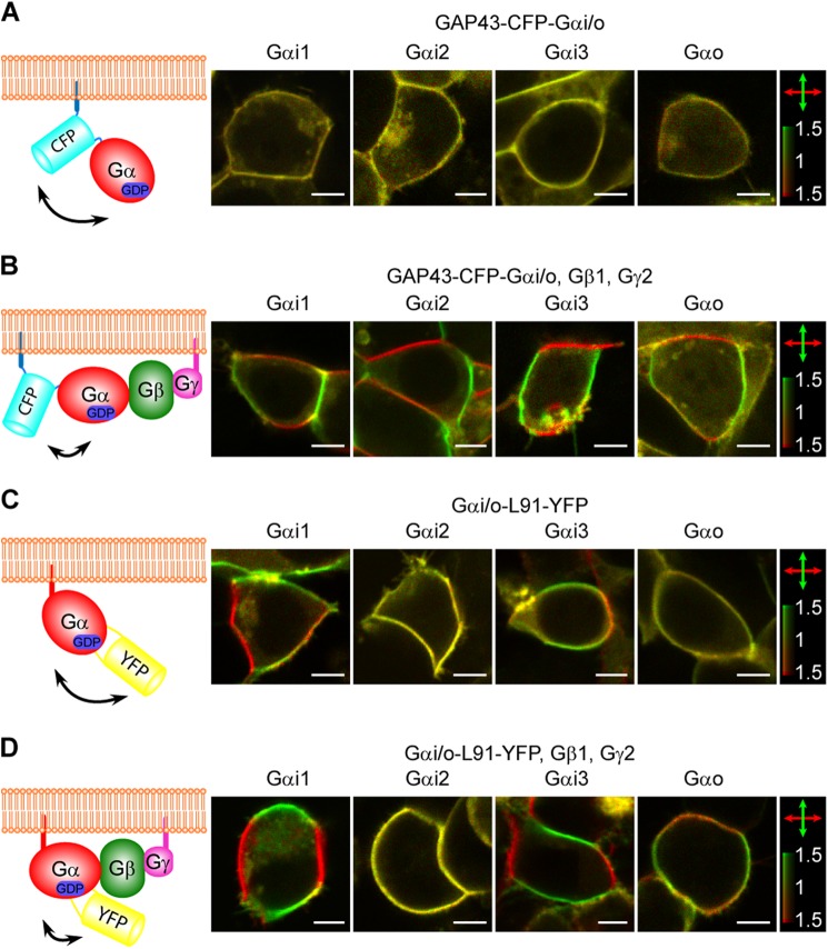
Imaging interactions between G protein subunits by 2PPM. A, 2PPM imaging of cells overexpressing Gαi/o-FP constructs of the GAP43-CFP-Gαi/o design. A schematic of the GAP43-CFP-Gαi/o construct design with high conformational flexibility expected (double-headed arrow). Also shown are 2PPM images of HEK293 cells transfected with individual GAP43-CFP-Gαi/o constructs. Scale bars = 5 μm. Fluorescence elicited by horizontal and vertical excitation beam polarizations is colored red and green, respectively (double-headed arrows), with the color bar indicating the dichroic ratio r. The absence of red or green color in the images indicates the absence of LD. B, same as A, but for GAP43-CFP-Gαi/o constructs coexpressed with Gβ1 and Gγ2 subunits. LD (a red/green pattern) is apparent in the outlines of cells expressing all four GAP43-CFP-Gαi/o constructs. C, same as A, but for Gαi/o-L91-YFP constructs. LD is present in cells expressing the Gαi1-L91-YFP and Gαi3-L91-YFP constructs but not the Gαi2-L91-YFP or Gαo-L91-YFP constructs. D, same as C, but the Gαi/o-L91-YFP constructs were coexpressed with Gβ1 and Gγ2 subunits. LD is present in cells expressing all but the Gαi2-L91-YFP construct. Interestingly, the distribution of fluorescence intensities (localization of red/green parts of the cell outline) indicates that although the YFP fluorophore in the Gαi1-L91-YFP and Gαi3-L91-YFP constructs is oriented close to perpendicular to the cell membrane, in the Gαo-L91-YFP construct it is close to being parallel to the cell membrane. In Gαi2-L91-YFP, the fluorophore is either in a disordered orientation or close to the magic angle, 52.0 degrees, to the cell membrane.

