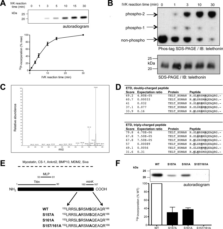
FIGURE 1.
PKD phosphorylates telethonin at Ser-157 and Ser-161. A, time course of PKD-mediated phosphorylation of WT telethonin. Recombinant WT telethonin was incubated with PKDcat and [32P]ATP and 32P incorporation over time was monitored by SDS-PAGE and autoradiography. Top panel, representative autoradiogram with quantitative data below (n = 4, mean ± S.E.). IVK, in vitro kinase. B, Phos-tag phosphate-affinity SDS-PAGE and immunoblot analysis of PKD-mediated phosphorylation of WT telethonin showing non-phosphorylated and phosphorylated moieties (top panel). Standard SDS-PAGE and immunoblot (IB) analysis of the same samples is also shown (bottom panel) (n = 3). C, collision-induced dissociation spectrum from the doubly charged, doubly phosphorylated telethonin fragment peptide showing the characteristic neutral losses (-49 and −98 Da) of two phosphate groups from the peptide. D, Mascot search results obtained for electron transfer dissociation (ETD) fragmentation spectra of the doubly and triply charged precursor. S, phosphorylated serine residue. E, schematic of telethonin, illustrating protein interactions reported previously. For interactions underscored by a dashed line, the telethonin domains involved have not been mapped. Ankrd2, ankyrin repeat domain-containing protein 2; BMP10, bone morphogenetic protein 10; CS-1, calsarcin 1; MDM2, murine double minute 2; MLP, muscle LIM protein. Also shown are amino acids 152–166 for all full-length recombinant telethonin proteins generated. F, representative autoradiogram showing PKD-mediated phosphorylation of recombinant WT and mutated telethonin proteins (top panel) with quantitative data below (n = 3, mean ± S.E.).

