FIGURE 3.
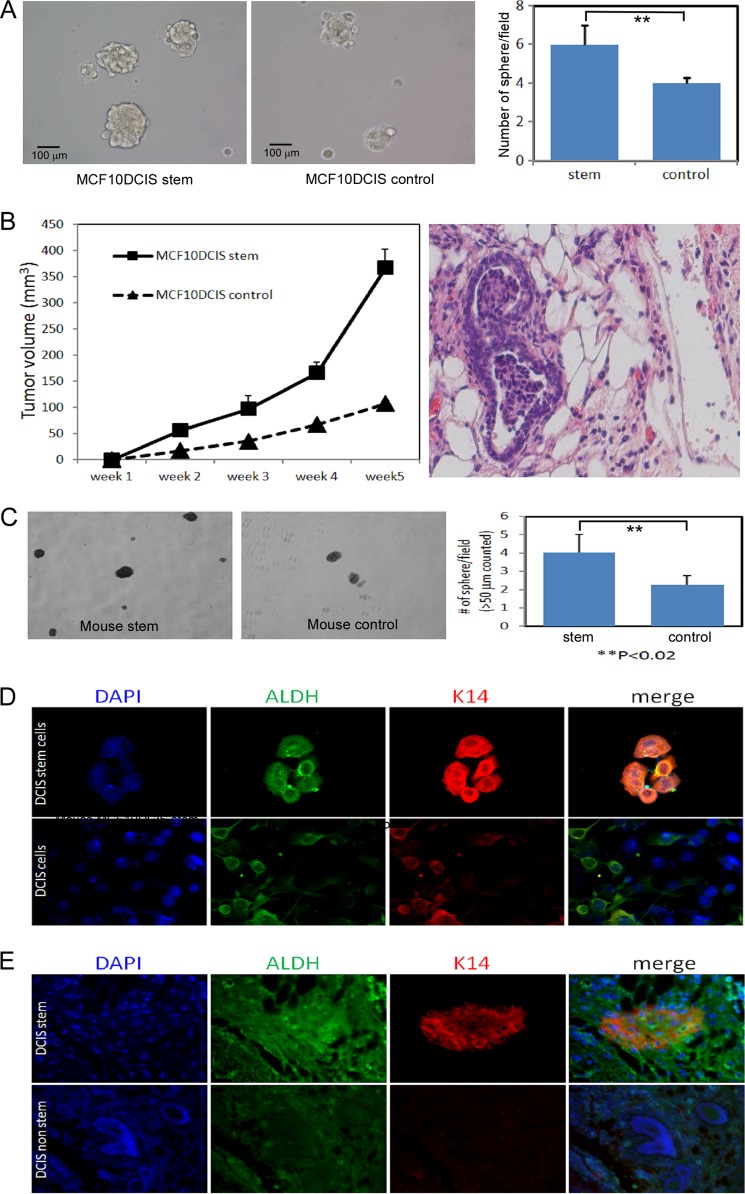
DCIS stem cells can form mammospheres and are enriched in ALDH1 staining in vivo. A, mammosphere formation of CD49f+/CD24− MCF10DCIS cells and MCF10DCIS control cells. B, DCIS stem-like cells are highly tumorigenic in vivo. Nude mice were injected with CD49f+/CD24− MCF10DCIS stem-like cells or MCF10DCIS control cells, and tumor growth was monitored weekly using digital calipers (left). Only one of five mice injected with MCF10DCIS control cells showed DCIS. However, four of five mice injected with MCF10DCIS stem cells showed DCIS. H&E staining of MCF10DCIS stem cell xenograft tumor showing DCIS structure (right). C, tumor cells were harvested and dissociated and then grown in attachment free mammosphere culture. D, DCIS stem-like cells were grown as mammosphere for 7 days after which cells were harvested and allowed to attach to coverslips for 24 h before performing immunofluorescence for ALDH1 and cytokeratin 14 (K14) and counterstained for DAPI. E, tissue from xenograft tumors (DCIS stem-like cells or DCIS non-stem cells) were examined by immunofluorescence for ALDH1 and cytokeratin 14.

