FIGURE 2.

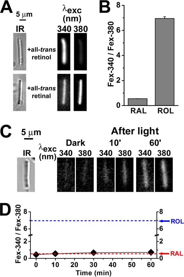
Measurement of RAL and ROL Fex-340/Fex-380 fluorescence ratios. Fluorescence was excited with 340- and 380-nm light, and emission was collected for >420 nm. A, fluorescence of mouse broken off rod outer segments (rod outer segments separated from the rest of the cell) loaded with 50 μm all-trans-retinol or all-trans-retinal (for 5 min, using 1% BSA as carrier). IR, infrared images of the outer segments. Fluorescence images of the outer segments after loading with retinoid are shown with the same intensity scaling to facilitate comparisons. B, values of the Fex-340/Fex-380 fluorescence intensity ratio for retinal and retinol, obtained by loading broken off rod outer segments with 50 μm RAL (n = 6 cells) and 50 μm ROL (n = 8 cells). C, mouse broken off rod outer segments cannot support the reduction of all-trans-retinal to retinol. IR, infrared image of an outer segment; fluorescence images of the outer segment before (Dark) and at different times after bleaching are shown with the same intensity scaling to facilitate comparisons; glucose concentration was 5 mm. D, dependence of the Fex-340/Fex-380 fluorescence intensity ratio on time after bleaching in broken off rod outer segments (♦, n = 6), in the absence of any exogenously added retinoid. The arrows point to the values of the ratio for RAL and ROL as determined in B. The value of the fluorescence ratio for the endogenous retinoid that appears after bleaching is similar to that for all-trans-retinal. All experiments were performed at 37 °C.
