
FIGURE 2.
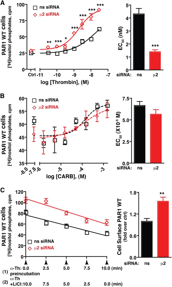
Activated PAR1 signaling efficiency, and not desensitization, is regulated by AP-2. HeLa cells expressing PAR1 WT were labeled with myo-[3H]inositol incubated with varying concentrations of thrombin for 5 min (A) or carbachol (CARB) or 10 min (B) at 37 °C. The concentration effect curve (mean ± S.D., n = 3) shown is a representative experiment. The EC50 values (mean ± S.E.) from multiple independent experiments are shown in the bar graph. The differences in thrombin-stimulated signaling were significant as determined by two-way ANOVA and Bonferroni post-tests (*, p < 0.05; **, p < 0.01; ***, p < 0.001). The differences in EC50 values were determined by Student's t test (***, p < 0.001). ns, not significant; Ctrl, control; M, molar. C, PAR1 WT-expressing HeLa cells labeled with myo-[3H]inositol were incubated with 10 nm thrombin for 10 min at 37 °C (1), and LiCl was added after various times of thrombin preincubation (2). [3H]IPs formed were then measured. The data (mean ± S.D., n = 3) are representative of three independent experiments. The differences in PAR1 surface expression (mean ± S.D., n = 3) were significant as determined by Student's t test (**, p < 0.01). The rates of desensitization (slope of the line) between nonspecific (-3.85 ± 0.43, n = 3) and μ2-adaptin-specific siRNA-treated cells (-4.76 ± 0.43, n = 3) were not significant as determined by Student's t test (p = 0.157). α-Th, thrombin.