
FIGURE 8.
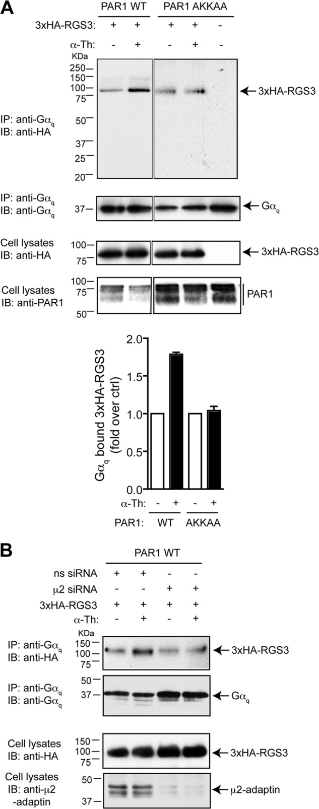
AP-2 mediates activated PAR1-stimulated Gαq-RGS protein complex formation. A, HeLa cells stably expressing FLAG-tagged PAR1 WT or AKKAA mutant were transiently transfected with HA-tagged RGS3 (3×HA-RGS3) and incubated with 10 nm thrombin (α-Th) for 5 min at 37 °C. Equivalent amounts of cells lysates were immunoprecipitated (IP) with anti-Gαq protein antibody, and coassociated RGS3 was detected with anti-HA antibody. Cell lysates were probed with anti-HA, anti-actin, and anti-PAR1 antibody as controls. IB, immunoblot. B, HeLa cells stably expressing FLAG- tagged PAR1 WT were transfected with nonspecific (ns) or μ2-adaptin-specific siRNA together with HA-tagged RGS3. Cells were then incubated with 10 nm thrombin for 5 min at 37 °C, and immunoprecipitated as described above. Cell lysates were immunoblotted with anti-μ2-adaptin antibody to evaluate the efficiency of AP-2 depletion. The data are representative of three independent experiments.