FIGURE 1.
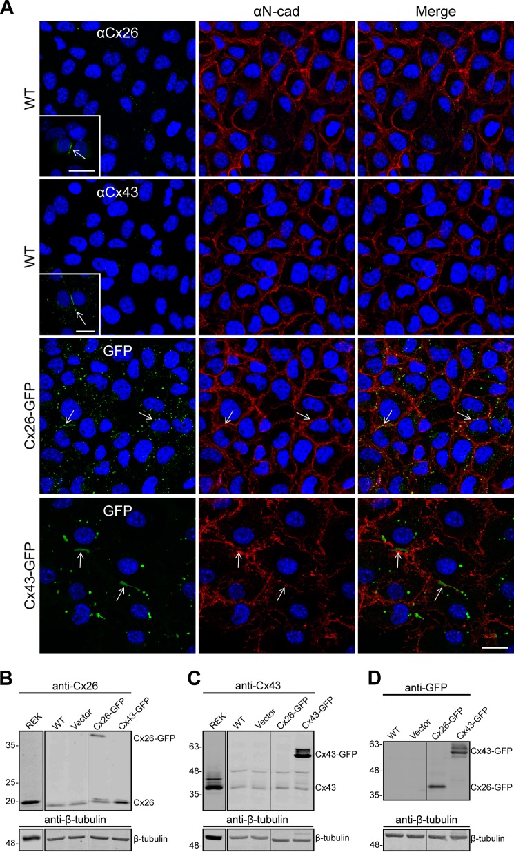
BL6 mouse melanomas express low levels of Cx26 and Cx43. A, immunofluorescence revealed that WT BL6 cells endogenously express low levels of Cx26 and Cx43 compared with REKs, which display typical gap junctional plaques at sites of cell-cell apposition (inserts). B and C, Western blots confirmed the low levels of Cx26 and Cx43 in melanomas compared with lysates collected from over-confluent and confluent REK cultures, respectively. Following ectopic expression of Cx26-GFP or Cx43-GFP, punctate gap junction-like plaques were evident at the cell surface as denoted by the cell adhesion molecule N-cadherin (red) (A, arrows), and the expression of GFP-tagged connexins was readily detected by Western blots immunolabeled for Cx26 (B) or Cx43 (C). D, additionally, both fusion proteins were expressed at similar levels as detected by Western blots immunolabeled for GFP. β-Tubulin was used as a loading control. The vector control represents cells transfected with a construct that did not encode connexins. Lanes separated by vertical lines were run on the same blot but spliced together. Lanes loaded with REK lysates were run as independent experiments. Blue, nuclei; bars, 20 μm.

