
FIGURE 2.
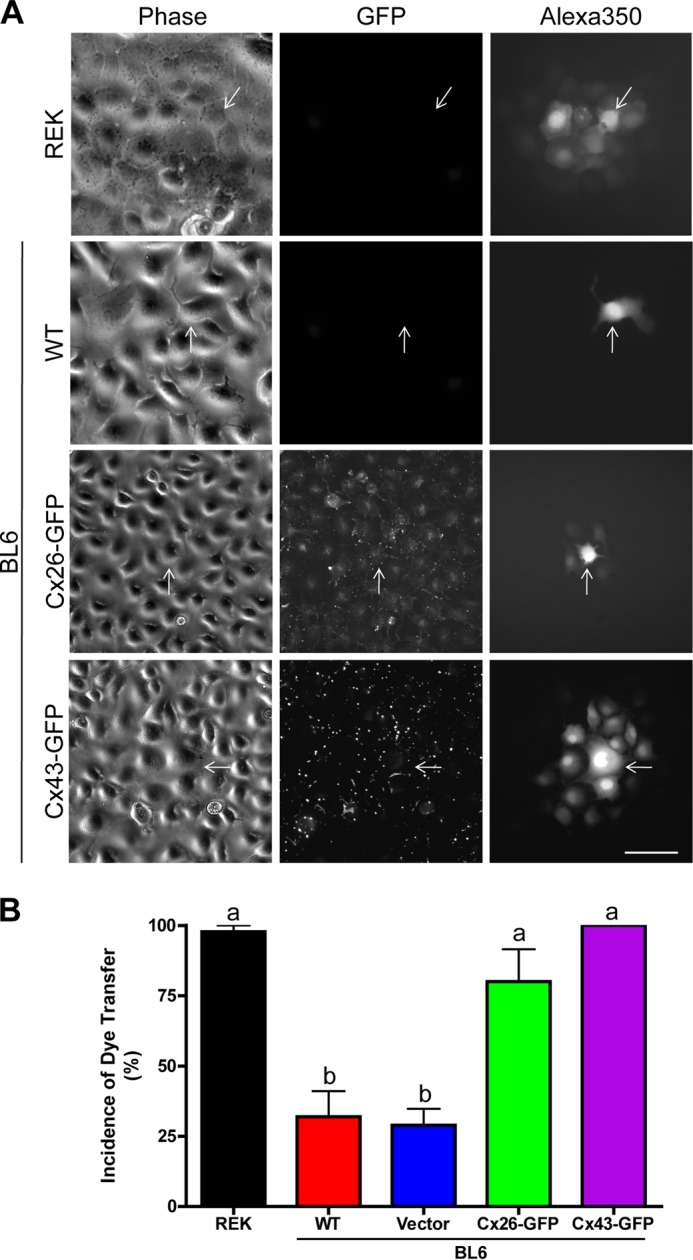
Ectopic connexin expression significantly increases GJIC. A, Alexa350 dye transfer studies revealed that wild-type and control melanomas were poorly coupled, because dye rarely spread from the microinjected cell (arrows) to neighboring cells. B, following ectopic expression of Cx26-GFP or Cx43-GFP, the incidence of dye transfer was significantly increased to 80 and 100%, respectively, statistically similar to Cx43-rich REK controls (n = 45, p < 0.05). Phase contrast images depict cellular morphology prior to microinjection, whereas the GFP fluorescence denotes the expression of ectopic connexins. Letters depict statistical significance among the groups. Bar, 40 μm.