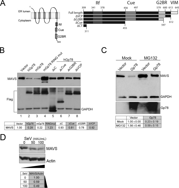
FIGURE 6.
The E3 ubiquitin ligase activity of Gp78 and its association with the ERAD pathway is required for Gp78-mediated MAVS degradation. A, left panel, schematic of Gp78 showing important regions for E3 ubiquitin ligase activity. Right panel, schematic of the C terminus of Gp78 illustrating deletion mutants used in this panel. B, immunoblot analysis from 293T cells 48 h post-transfection with EGFP-MAVS and the indicated plasmids. Antibody directed against GFP was used. Immunoblotting for Flag and GAPDH (bottom panel) are included to show expression of wild-type and mutant Gp78 and as a loading control, respectively. Table at bottom, densitometry (MAVS/GAPDH normalized to vector control) in cells transfected with the indicated plasmids. Densitometry was performed, and data are presented as fold change from vector untreated (bottom panel). C, immunoblot analysis from 293T cells 48 h post-transfection with EGFP-MAVS and either vector control or Gp78. MG132 (20 μm) was added 16 h post-transfection. Antibodies directed against GFP and GAPDH were used. Table at bottom, densitometry (MAVS/GAPDH normalized to vector control) in cells transfected with the indicated plasmids and either Mock- or MG132-treated. D, immunoblot analysis of MAVS (top) from 293T cells infected with SeV (50 or 100 HAU/ml) for ∼24 h. Immunoblotting for actin (bottom) is included as a loading control. Table at bottom, densitometry (MAVS/Actin normalized to control (mock infection).

