
Table 1.
Ultrasonographic and Doppler features used to evaluate liver cirrhosis
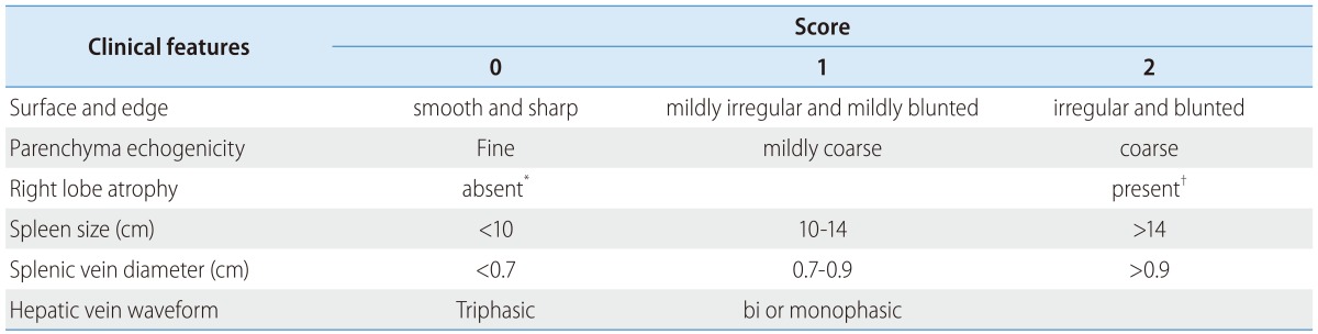
The total score from six ultrasonographic indices including surface nodularity and edge shape (0-2), parenchyma echogenicity (0-2), right lobe atrophy (0-2), spleen size (0-2), splenic vein diameter (0-2) and hepatic vein waveform (0-1) was calculated.
*Right lobe maximal oblique diameter >7 cm with no subphrenic ascites.
†Right lobe maximal oblique diameter <10 cm with subphrenic ascites.