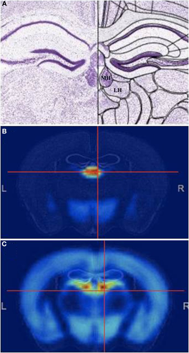
Figure 1.

The Medial habenula. (A) Mouse coronal brain section at the level of the medial habenula, stained with Nissl. Medial (MHb) and lateral (LHb) are indicated. (B,C) Pattern of general genetic co-expression in the mouse brain using the MHb (B) or the LHb (C) as seeds. Note that the expression patterns are completely different. All figures from the Allen Brain Atlas (Lein et al., 2007).
