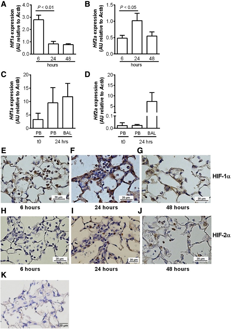
Figure 5.
Differential regulation of HIF1 and HIF2 in circulating neutrophils and neutrophils recruited to the lungs following LPS-induced lung injury. (A-D) C57BL/6 mice were instilled with intratracheal LPS (0.3 µg). BAL was performed at 6, 24, and 48 hours. (A) Hif1a and (B) Hif2a expression in BAL cell lysates from C57BL/6 mice determined by Taqman and normalized to Actb. (C) Hif1a and (D) Hif2a expression in freshly isolated (t0) peripheral blood neutrophils (PBs), or peripheral blood neutrophils and BAL cells isolated 24 hours after LPS instillation. Data are mean and SEM for n = 3. (E-J) Immunohistochemistry showing expression of (E-G) HIF-1α and (H-J) HIF-2α in neutrophils of WT mice (E,H) 6, (F,I) 24, and (G,J) 48 hours following challenge with nebulized LPS (3 mg). (K) Immunohistochemistry showing no HIF-2α expression in myeloid-specific HIF-2α–deficient mouse lungs. Original magnification ×400. Images were taken using a Nikon Eclipse E600 microscope, with a Nikon DS-Ri1 camera, and processed with NIS-Elements Basic Research software (Nikon, Kingston upon Thames, United Kingdom).

