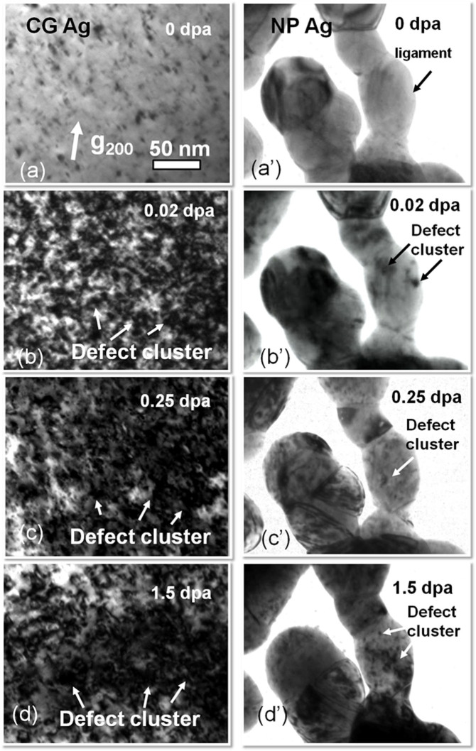
Figure 1. Drastic difference between microstructures of CG (a–d) and NP (a′–d′) Ag subjected to in situ Kr ion irradiation at room temperature.
(a-a′) Annealed CG Ag films had a low density of preexisting defects, whereas NP Ag was nearly defect free. (b-b′) After irradiation to 0.02 dpa, CG Ag was swamped with a large number of defect clusters, whereas NP Ag remained intact (see Supplementary Video 2). (c-c′) By 0.25 dpa, there was a significant increase in both the size and density of defect clusters in CG Ag. In parallel few defect clusters were observed in the ligaments of NP Ag. (d-d′) By 1.5 dpa, the average defect cluster size in CG Ag appeared much greater than that in NP Ag.

