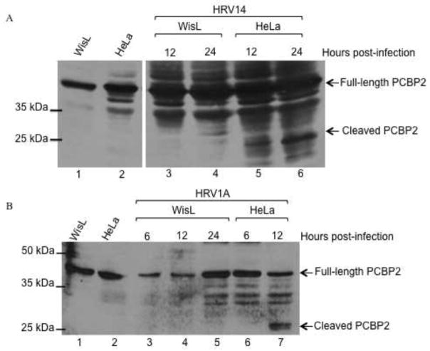
Fig. 6. PCBP2 cleavage during human rhinovirus infection.
HeLa or WisL cells were infected with HRV and cytoplasmic lysates were collected at indicated times post-infection. The collected lysates were then analyzed by Western blot with an antibody specific for PCBP2. (A) Cells were infected with HRV14. Extracts from mock-infected WisL or HeLa cells are shown in lanes 1 and 2, respectively. Lysates from infected HeLa cells are shown in lanes 3 and 4, and lysates from infected WisL cells are analyzed in lanes 5 and 6. (B) WisL (lanes 3–5) or HeLa cells (lanes 6 and 7) were infected with HRV1A. Extracts from mock-infected WisL or HeLa cells are analyzed in lanes 1 or 2, respectively. Arrows on the right of the gel image indicate full-length or cleaved PCBP2, and molecular weight markers are on the left of the gel image.

