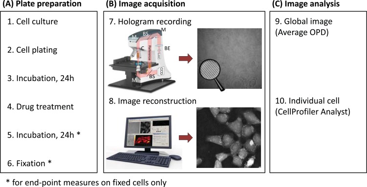
Fig. (1).
The Digital Holographic Microscopy (DHM) technology and experimental workflow. (A) Plate preparation procedure. (B) Image acquisition: First, a hologram (magnifying glass highlights the interference fringes) is recorded out of focus by a digital camera on a DHM T1001 system equipped with a motorized stage for automated multi-well plate experiments (7). Legend: M, mirror, BS, beam splitter, BE, beam expander, MO, microscope objective, C, condenser. Then, it is reconstructed by a computer to form an in-focus quantitative phase image (8). Contrast in DHM is provided by optical path length (OPL) variations in the specimen. For cell biology experiments, the measured optical path difference (OPD) is related to the thickness d and mean intracellular refractive index nc of the cultured cells, as well as to nm, the refractive index of the surrounding medium through Equation (1). (C) Image analysis is performed on the quantitative phase images using either on the global image (9) or on individual cell (10).

