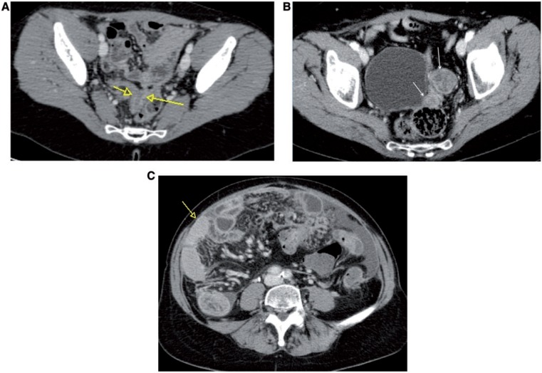
Figure 22.
Axial CT images showing different appearances of bowel infiltration by ovarian cancer: inhomogeneous enhancement of the bowel serosa (A); presence of enhancing nodular masses within the left pelvis (arrows) infiltrating the distal part of the large bowel (B); presence of abnormal mural thickening of small bowel (arrow) (C).

