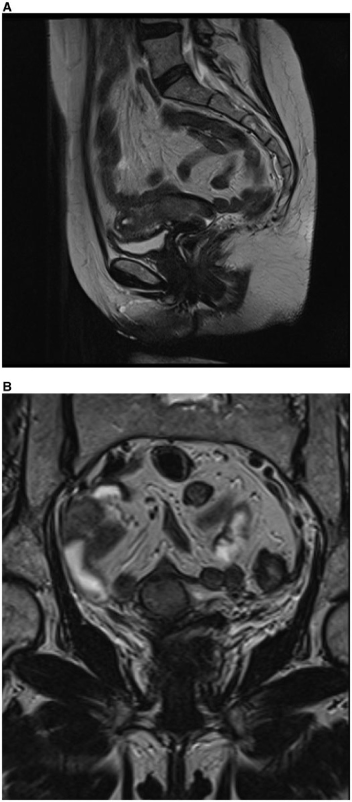
Figure 5.
Sagittal (A) and para-axial (B) MR T2-weighted images showing endometrial cancer extending to the cervix, with preserved hypointense stromal ring, as confirmed by regular enhancement at dynamic postcontrast T1-weighted image (C) in an endometrial carcinoma, International Federation of Gynecology and Obstetrics (FIGO) stage IIA.

