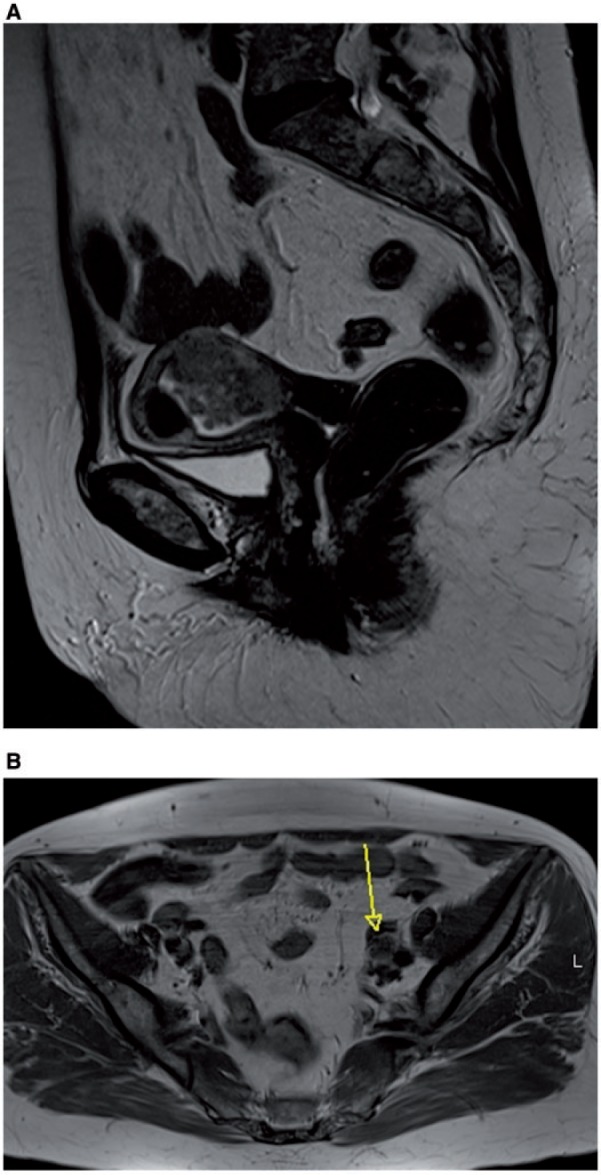
Figure 6.

Sagittal MR T2-weighted image showing endometrial cancer in the middle part of the uterine corpus, invading the outer part of myometrium (A), which led to nodal metastasis to obturator left lymph node (arrow), as shown in the axial unenhanced MR T1-weighted image (B).
