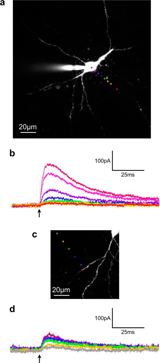
Figure 2.

Spatial resolution of one-photon uncaging of compound 6 with 473 nm light. Compound 6 was bath applied at 10 μM to an acutely isolated brain slice and photolyzed (arrows) with a continuous wave, 473 nm laser (1 ms, 2mW) centered at the indicated points. (a) Two-photon fluorescence image of a CA1 pyramidal neuron with the positions of uncaging marked. (b) The outward current traces corresponding to each point in (a). (c) Two-photon fluorescence image of a CA1 pyramidal neuron filled with Alexa-594 with the positions of uncaging marked on or near an isolated dendrite (d) The outward current traces corresponding to each point in (c). Cells filled with Alexa-594 were imaged at 1075 nm.
