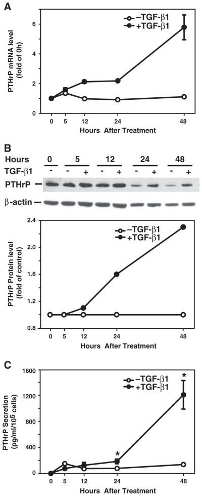
Fig. 1.
TGF-β induces PTHrP expression and secretion. Hep3B cells were cultured in the presence or absence of TGF-β1 (40 pmol/L) over a 48 h time course. A. Total RNA was prepared at 0, 5, 12, 24, and 48 h after TGF-β1 treatment, and converted to cDNA. PTHrP mRNA expression was detected by real-time quantitative PCR. PTHrP mRNA level was expressed as fold of the value at time 0 h. B. Cell lysates were prepared for Western blotting using specific antibody against PTHrP. β-actin served as a protein loading control. Western blotting images are presented on the top panel and PTHrP protein levels were normalized to β-actin and presented as folds of time-matched controls without TGF-β1 on the bottom panel. C. Cell culture supernatants were collected for measurement of PTHrP peptide secretion. The concentration of PTHrP from triplicate wells is expressed as pg/ml and normalized by cell number. Results from triplicate wells are expressed as mean ± SEM. *p < 0.05 compared to the group in the absence of TGF-β1.

