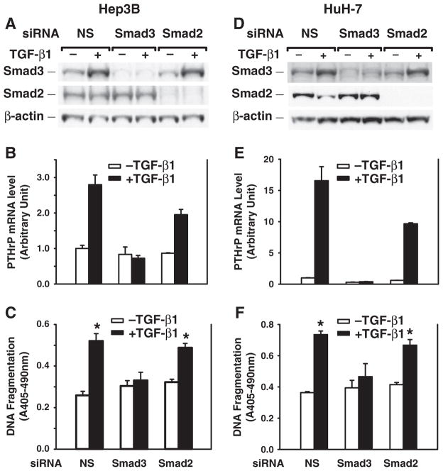
Fig. 6.
Silencing Smad3 gene expression blocks TGF-β-induced PTHrP expression and apoptosis induction. Hep3B and HuH-7 cells were transfected with human Smad3 or Smad2 siRNA (100 nmol/L) using DharmaFECT2 and then replated 24 h after siRNA transfection. Cells were then cultured in the presence or absence of TGF-β1 (40 pmol/L) in MEM supplemented with 0.1% FBS for 48 h. A, D. Whole cell lysates were prepared for Western blotting of Smad3 and Smad2. B, E. Total RNAs were prepared and converted to cDNAs for real-time quantitative PCR analysis of PTHrP mRNA expression. C, F. DNA fragmentation was quantified by cell death detection assay. Results from triplicate wells are expressed as mean ± SEM. *p < 0.05 compared to the group in the absence of TGF-β1.

