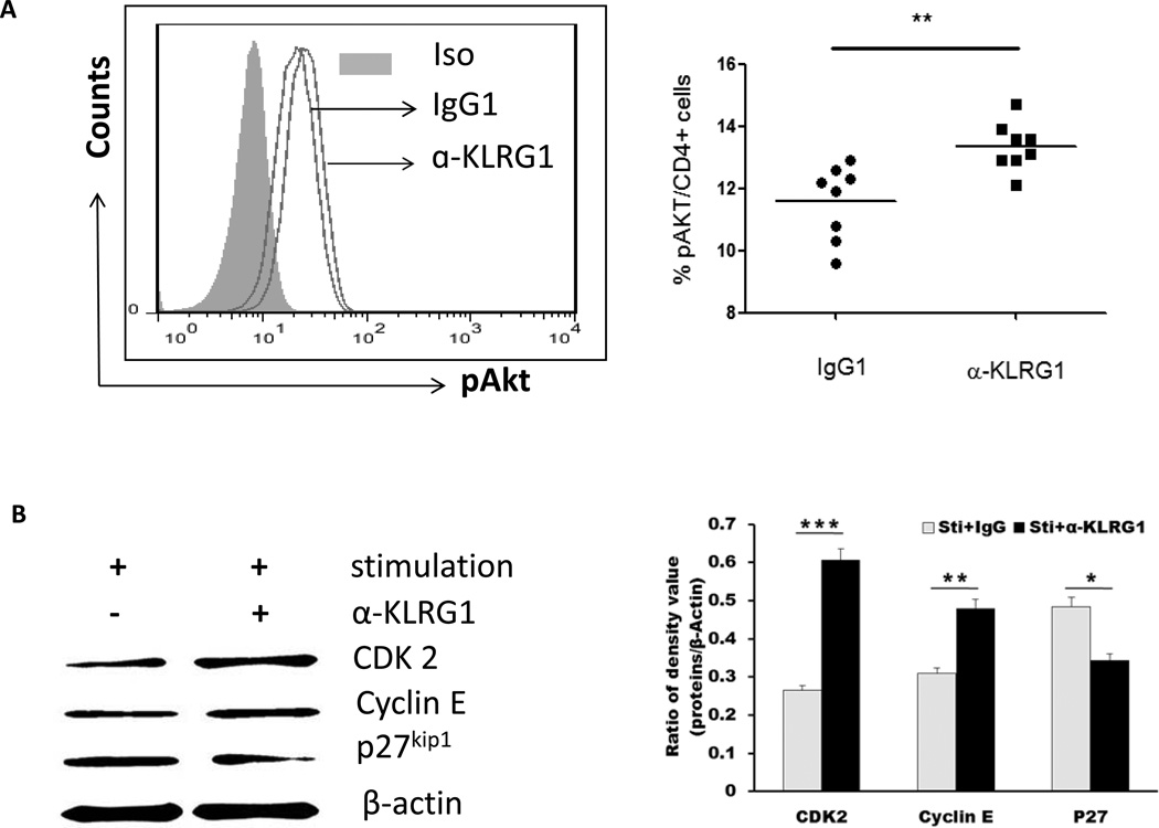
Fig. 5. KLRG1 inhibits Akt (Ser473) phosphorylation and downstream signaling pathways in CD4+ T cells during HCV infection.
A) KLRG1 blockade increases pAkt (ser473) phosphorylation in purified CD4+ T cells from HCV-infected HBV-NR. Left panel: representative histograms of pAkt (ser473) expression in the CD4+ T cells treated with control IgG1 or anti-KLRG1. Grey-filled area is isotype control staining. Right panel: summary of the percentages of pAkt (ser473) phosphorylation in CD4+ T cells from HBV-NR following treatment with anti-KLRG1 or control IgG. The horizontal bars indicate the median values for 8 HCV infected HBV-NR. **P<0.01. B) T cells of PBMCs from HCV-infected HBV-NR were stimulated by anti-CD3/CD28 in the presence of anti-KLRG1 or control IgG for 3 days, and total protein were extracted from the cell lysates, followed by Western blot analysis of CDK2, Cyclin E and p27kip1 expressions. β-actin serves as loading control. Representative imaging are shown in the left panel, and summary densitometry data of CDK2, Cyclin E and p27kip1 expressions, corrected by β-actin level, from three independent experiments are shown in the right panel. *P<0.05; **P<0.01; ***P<0.001.

