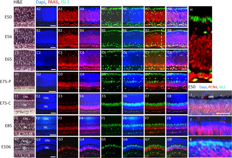
Figure 1.
Postmitotic PAX6−, ISL1+ cells comprise the outer rows of the retina before midgestation in the pig. (A–G) H&E and immunostaining of swine retinal sections from the indicated embryonic days. The boxed region in (B7) is shown in (H). (I–K) Co-immunostaining for PCNA and ISL1 at E50. (I) Nomarski image showing immunostaining for PCNA. (J) DAPI and PCNA immunostaining. (K) Double immunostaining for PCNA and ISL1 along with DAPI. C, central retina; P, peripheral retina (see Materials and Methods). Scale bars: 50 μm.

