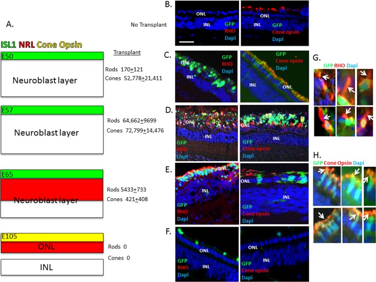
Figure 7.
Evaluation of integration/differentiation following transplantation of embryonic retinal cells into the subretinal space of adult pigs. (A) Summary of the time course of appearance of cone precursors (green; marked by transient expression of ISL1 and early expression of RCVRN) and rod precursors (red; marked by NRL) in the developing pig retina. (B) No transplant shows loss of RHO expression and diminished expression of cone L/M opsin in retinas after IAA treatment (Materials and Methods; Ref. 16). (C–F) Representative examples of immunostaining of GFP+ transplanted cells for cone opsin and RHO is shown corresponding to embryonic ages in (A). The average number ± SEM of integrated GFP/RHO+ (rods) and GFP/cone opsin+ (cones) in at least three independent injections of 1,000,000 cells is shown. GFP+ integrated cells were counted in 20-μm frozen sections across the injection site, which corresponded in size to approximately 2 optic disc diameters. (G, H) High-power views showing colocalization of GFP and RHO or cone opsin. Arrows indicate OS. Scale bar: 50 μm. P < 0.01 for rod number at E50, E57, and E65, and for cone number at E50 and E57 versus E65.

