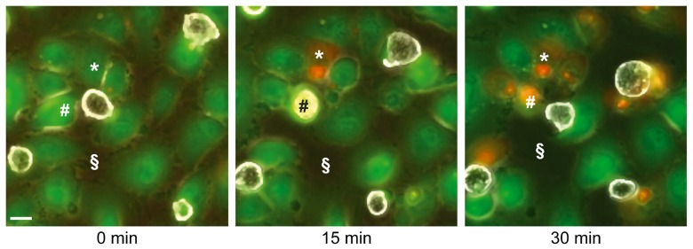
Fig. 3.
Retraction and cell death in LSEC cultures during incubation with E. histolytica. Micrographs of video-microscopy sequences show frames taken at 0, 15 and 30 min of incubation. LSEC were labelled with fluorescent CMFDA cell tracker (green). Trophozoites were then added (parasite to LSEC ratio 1:10) and observed by phase contrast microscopy, as non-fluorescent cells (white) with brightly reflecting plasma membranes. Dead cells were detected by incorporation of propidium iodide (red). Note that human cells (i.e. * and #) were in contact with an amoeba before dying. The space not occupied by cells (i.e. §) is increasing over time, indicating LSEC retraction. Scale bar, 10 µm

