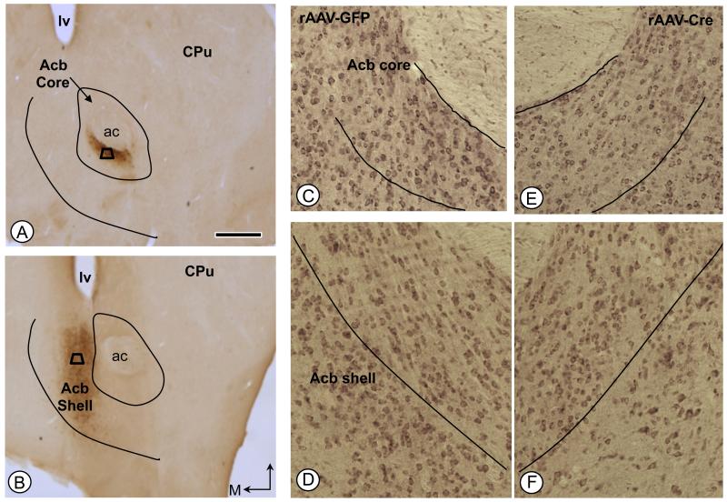
Figure 2.
Immunoperoxidase labeling of the GFP reporter protein in the Acb following local unilateral microinjection of vector into either the core (A) or shell (B) subregions. Areas bounded by the trapezoids represent regions where tissue was sampled by EM in adjacent sections processed for dual D1R and μ-OR immunolabeling. (C-F). NR1 immunolabeling in the Acb core (C, E) and shell (D, F) of rAAV-GFP and rAAV-Cre injected mice, respectively. Bar=500 μm ac: anterior commissure, CPu: caudate-putamen, D: dorsal, lv: lateral ventricle; M: medial.

