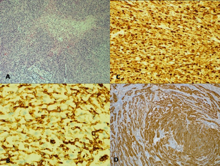
Figure 3.
Histological photograph of the tumour. (A) Gastric tumor showing necrosis (Haematoxylin and Eosin stain 10×), (B) CD34 immunostain is positive (20×), (C) S100 immunostain is positive (10×), (D) Immunohistochemistry picture showing positivity of the tumor cells to S100 indicating its neural origin.

