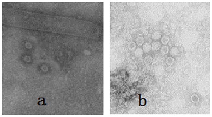
Figure 3. Electron microscopy of CPV-VLP.
Sample were negatively stained with 2% uranyl acete and observed by electron microscopy. A, Analysis of the particles formed in recombinant baculovirus infected silkworm pupae. B, Particles collected from sucrose gradients (purified VLP). Magnification 40,000×.

