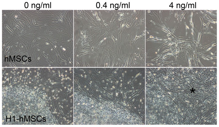
Figure 1. Morphological changes of hMSCs feeder cells and H1 cells in media supplemented with 0, 0.4 or 4/ml bFGF.

Upper row: hMSCs feeder cells presented distinct morphologies in media supplemented with 0, 0.4 or 4 ng/ml bFGF. Bottom row: H1-4 ng culture differentiated into short spindle-shaped fibroblast like cells at P32+8 as indicated by the asterisk (*). See also Figure S1.
