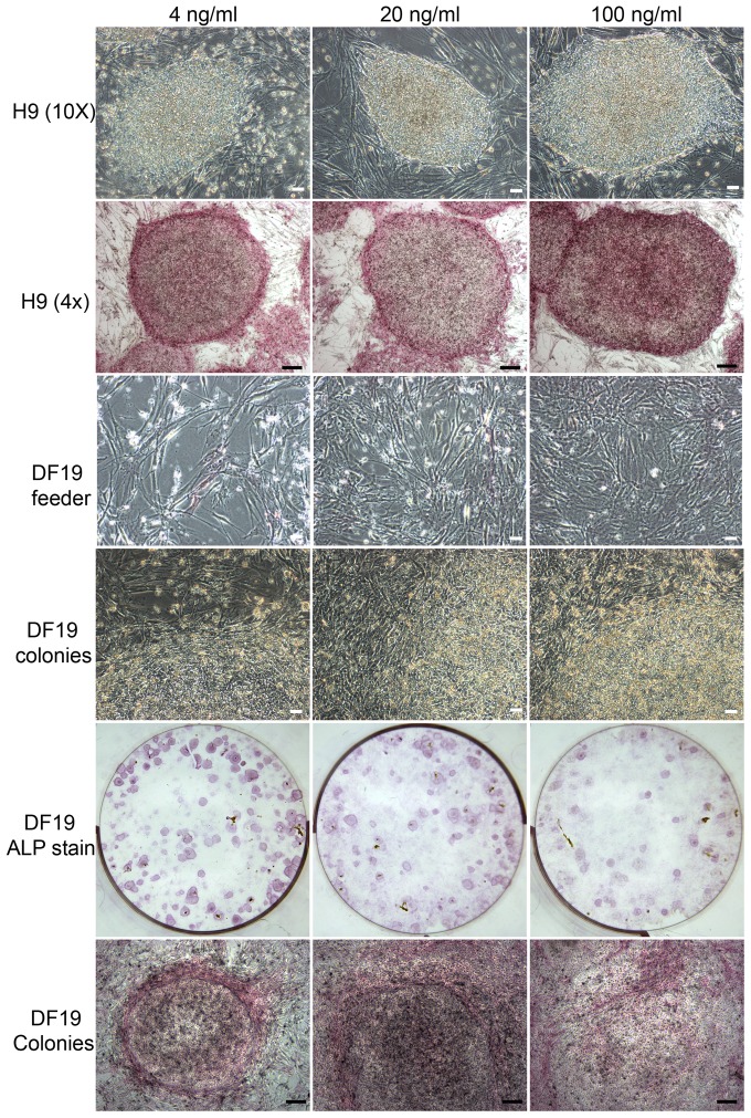
Figure 10. bFGF at 20 or 100/ml promoted DF19 differentiation in a dosage dependent manner but had limited effect on H9 cells.
Top two rows: Bright-field images and ALP stained images of H9-4 ng, H9-20 ng and H9-100 ng cultured in parallel at P40+11; Middle two rows: Bright-field images of feeder cell layers and colonies in DF19-4 ng, DF19-20 ng and DF19-100 ng cultures at P33+6, with the latter demonstrating more blurred boundaries between feeder cells and colonies in DF19-20 ng and DF19-100 ng; Bottom two rows: ALP stained images of DF19-4 ng, DF19-20 ng and DF19-100 ng cultures at P33+6. Scales in white: 50 µM; Scales in black: 200 µM.

