Figure 3:

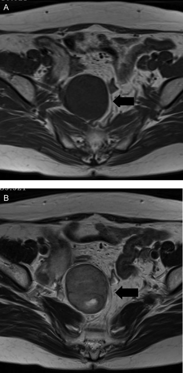
MRI shows that the tumor is a homogeneous hypointensity on T1-weighted images (A) and a heterogeneous slight hyperintensity on T2-weighted images (B) in a right side of the pelvic cavity.

MRI shows that the tumor is a homogeneous hypointensity on T1-weighted images (A) and a heterogeneous slight hyperintensity on T2-weighted images (B) in a right side of the pelvic cavity.