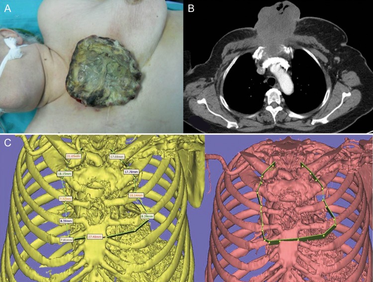
Figure 1:
Preoperative view of the chest wall tumour. The mass involves the skin and anterior chest wall including manubrium sterni (A). The preoperative computed tomographic scan of the patient reveals no invasion to the vascular structures of the mediastinum. It also shows that most parts of the anterior chest wall bone structures were invaded by the mass (B). The resection plan was marked on the 3D reconstruction of the computed tomography scan (C).

