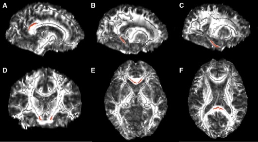
Figure 1.

White matter ROIs (shown in red) overlayed on the FA template image (skeletonized FA underlayed in light gray). The bilateral ROIs included: (A) cingulum bundle subjacent to posterior cingulate (119 voxels, MNI coordinates: ±11,−45, 28), (B) cingulum adjacent to hippocampus (60 voxels, MNI coordinates: ±20, −42, −2), (C) entorhinal white matter (96 voxels, MNI coordinates: ±24, −26, −19), (D) corticospinal tract (31 voxels, MNI coordinates: ±10, −20, −24), (E) splenium (49 voxels, MNI coordinates: ±1, −35, 14), and (F) genu (48 voxels, MNI coordinates: ±4, 23, −1) of the corpus callosum. (A–C) Sagittal view, (D) coronal view, and (E, F) an axial view.
