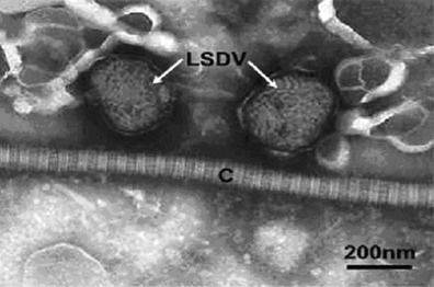
Fig. 4.

Transmission electron micrograph of two negatively PTA-stained LSDV particles indicated (arrows) in close association with a collagen fibre (C). The particles show a typical thread-like structure on their surface and typical “brick-shaped” morphology (arrows)
