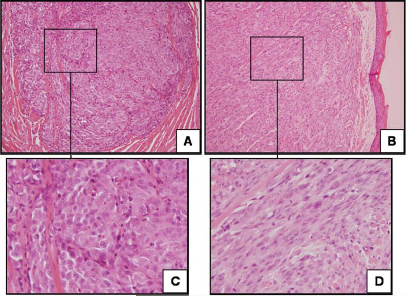
Fig. 3.
Xenograft histology from orthotopically established tumors. Representative histologic sections of xenografts established from UMSCC-74A treated with control (A and C) or recombinant human bone morphogenetic protein-2 (B and D). The xenografts showed changes in morphology toward more poorly differentiated tumors with spindle cell features. Hematoxylin and eosin; original magnification, 200 (A and B) and 400× (C and D).

