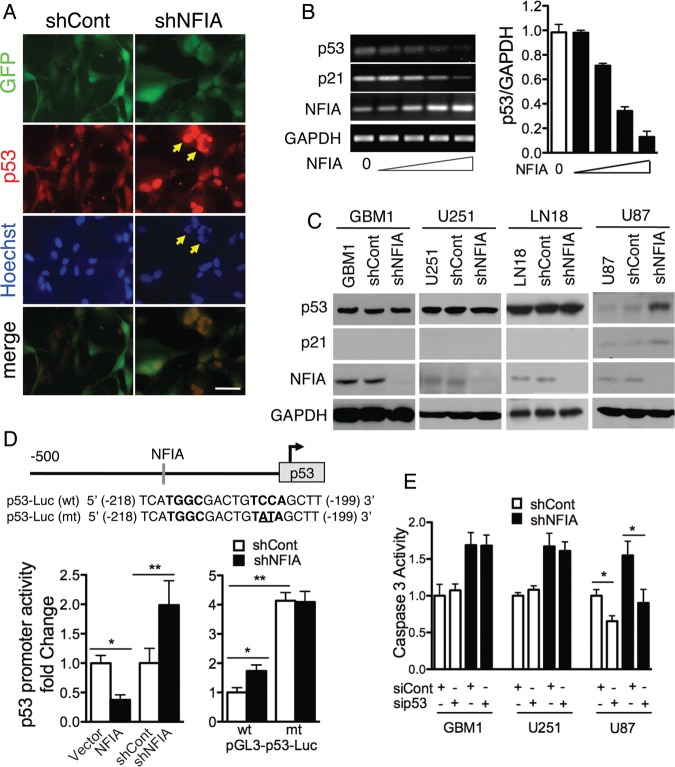
Fig. 3.
Apoptosis induced by loss of NFIA is mediated by p53. (A) Immunofluorescence of U87 cells expressing shNFIA and control (shCont) 4 days after transduction; p53 - red, Hoechst - blue. Arrowheads: examples of fragmented nuclei with high nuclear p53. Scale bar: 50 µm. (B) NFIA represses p53 mRNA. p53, p21, and NFIA mRNA (RT-PCR) from U87 cells transduced for 2 days with 0, 1/27, 1/9, 1/3, 1 µg lentiviral plasmid containing NFIA cDNA and adjusted to total 1 µg transfected DNA using empty-vector plasmid. Right panel: p53 mRNA normalized to GAPDH; n = 3; P < .0001 by 1-way ANOVA. (C) Loss of NFIA induces p53 in p53-wild-type U87 cells but not in the p53-mutant GBM1, U251 and LN18 cells. Glioma cells 3 days post lentiviral transduction with shNFIA or control shRNA were assessed for p53 and p21 expression by immunoblotting. (D) NFIA negatively regulates the p53 promoter. Top: NFIA consensus-binding sequence (−199 bp) in the p53 promoter. Left: relative luciferase activity 48 hours after transfection of p53 luciferase reporter into U87 cells stably expressing NFIA or vector or 3 days post lentiviral transduction with shNFIA or controls; *P < .005, **P < .05, see also Figure S4A. Right: relative luciferase assay using a wild-type p53 reporter or a mutated counterpart with destroyed NFIA binding site (see top panel) in U87 cells 3 days post lentiviral transduction with shNFIA or shCont; n = 3; *P < .01, **P < .001. (E) Knockdown of p53 attenuates shNFIA-induced caspase-3 activity in U87 cells but not in p53-mutant cells (GBM1 and U251). Glioma cells 3 days post lentiviral transduction with shNFIA or shCont were treated with p53 siRNA or control siRNA (100 nM) for 48 hours, and caspase-3 activity was measured; n = 3, *P < .05; Representative protein expression and densitometric analysis are shown in Figure S5.

